RONTGEN RAY (X-RAY) PHOTOGRAPHY
In November 1895 Professor Wilhelm Conrad Röntgen of Bavaria discovered the effect that rays from a Crookes tube had on a fluorescent screen, and this led to him discovering the effect they had on photographic plates. Because there was no explanation for the existence of these invisible rays Röntgen called them X-rays – ‘X’ denoting the unknown quantity. It was 15 years before the correct explanation for the rays was put forward.
On 22 December 1895 Röntgen used X-rays to photograph his wife’s hand, the world’s first X-ray photograph. Before the term radiograph came into general use, photographs taken by Röntgen rays were called skiagraphs or skiograms. When the news of the discovery reached Australia in January 1896 there were some who dismissed it as impossible, including the Australsian Photo-Review, which said ‘nobody appears to know who Röntgen is, or to have heard of him before.’ The Review could not come to terms with the idea of invisible light, and described the report from overseas as ‘misleading and delusive.’
Early in 1896 Professor Thomas Lyle of the Melbourne University was experimenting with the ‘new non-luminous rays’ and would have been the first person in Australia to make an ‘x ray’ photograph. In one experiment he produced a ‘shadow photograph’ of a human hand on a photographic plate after a twenty minute exposure to the rays, and told a reporter he would pursue his investigations but ‘could give no indication as yet of the practical value to the world of the new discovery.’
In May 1896 Mr S. Barbour, manufacturing chemist for F.H. Faulding & Co., returned from a holiday in Europe and America and brought with him two Crookes tubes. As the rays were a new discovery the tubes were in great demand and difficult to obtain, but he managed to obtain two from the makers, Reynolds and Branson of Leeds, which had been his home town before he came to live in South Australia.
Soon after he arrived in Adelaide Mr Barbour, assisted by Mr Rowe, began experimenting with the tubes, and he was the first person in South Australia to apply Röntgen rays to anatomical subjects. He was using half-hour exposures but thought that with a more powerful battery he might be able to bring this down to ten minutes. He tried the commercial power supply but the results were not satisfactory. On Thursday, 28 May, he secured a fair impression of a human hand, the outline of the bones being distinct, and photographed some objects through wood – a pair of scissors, a Maltese Cross, a triangle made of wire and covered with pipeclay, and an india-rubber ring. The scissors and cross were distinct, the ring was faint, and the wire triangle could be seen inside the pipeclay.
On Friday evening, 29 May, Barbour took the tubes to the University of Adelaide laboratory where a more powerful battery was available. The experiments were conducted by Professor Bragg of the University assisted by Mr Barbour, with Mr Rowe processing the negatives. Witnessing the experiment were Doctors Giles, Swift and London. The first experiment was was a five-minute exposure of Professor Bragg’s hand with a coin under one finger, the coin coming out as a black spot and all the bones being shown. Next was a photo of a foot taken to expose a defect in one toe, then a 25-minute exposure of a kneecap. The last experiment was a five-minute exposure of the wrist of one of the doctors, which came out ‘clear and distinct.’ The medical men were ‘highly delighted with the achievements’ and satisfied the discovery would be of great value to their profession. ‘Already there are people waiting who want the situation of needles and bullets in various parts of the body detected.’
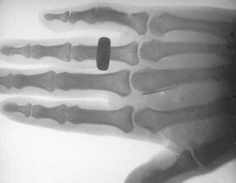
The newspaper report of the Röntgen ray experiments at the University aroused widespread interest among the members of the medical profession, so on Monday 1 June further experiments were conducted at the University in the presence of a large group of doctors and better results were obtained. While a 30-minute exposure to the rays gave a very clear picture of a foot encased in plaster of Paris produced a very clear picture, another experiment produced practical results. A lady who had been troubled by a piece of a needle lodged in her hand had ‘the afflicted member’ subjected to the rays. Although doctors had not been able to locate the piece of needle, in the photograph it could be clearly seen at the base of her third finger and only a simple operation was needed to remove it.’ (see copy of the X-ray below).
Below: Lantern slide copy of the X-ray referred to in the above paragraph. The mask on the slide bears the hand-written inked inscription, "Needle in hand for 5 years 2/9/96" and also the wet-stamp impression, "W.T. ROWE." The exposure could have been from 5 to 10 minutes with Mr S. Barbour of F.H. Faulding operating the apparatus and Mr W.T. Rowe developing the plate.
Professor Bragg had been experimenting with the rays for some time but had been handicapped through not having the proper tubes, and although the instrument fitter at the University, Mr A.L. Rogers, had managed to manufacture one tube it failed after one photograph was taken. Professor Bragg experimented with the manufacture of an opalescent screen which would glow in the dark and allow him to see the image without the need for photographs.
On Wednesday, 3 June, the Governor’s wife, Lady Buxton, and some of her children paid a visit to the University to see the Röntgen ray apparatus at work. ‘Some of the visitors submitted their hands to be photographed, while several little things belonging to Lady Victoria were placed in a matchbox and the contents photographed through the wood.’
On 17 June 1896 Professor Bragg demonstrated the effect of Röntgen rays to a large and ‘fashionable’ audience at the Adelaide University Library which included the Governor and his family, University officials, graduates, students and ‘prominent citizens and their ladies.’ With the aid of a sketch projected on a ‘huge’ screen the Professor described the production of Röntgen rays and their effect on photographic plates then proceeded with the demonstration.
‘Various opaque objects, some of them enclosed in cardboard boxes, were placed upon photographic plates, sheltered from the gaslight by many thicknesses of orange paper. These were on a small table, and were subjected to the effect of the Röntgen rays. At the same time, but exactly under the tube that was used, Mr Moir, department manager at Messrs. Faulding & Co.’s, placed his right hand on a cardboard box containing a large photographic plate. The exposure lasted about 8 minutes… The plates were then taken in hand for reproduction as lantern slides by Messrs. S. Barbour and W.T. Rowe…
‘The Professor referred to the efforts of Mr A.L. Rogers, of the University, in the making of a Röntgen tube. Only a few days ago their endeavours were crowned with success, and a photograph was taken, but soon afterwards the tube failed. A photograph, that of a frog, which was reproduced on the screen, was interesting as being the first [?] and only skiagraph obtained by means of an Australian-made tube. [Professor Lyle had made Röntgen ray photographs several months before this with a ‘Crookes’ tube he had made himself.]
‘Professor Bragg described the successful working of the tube [he was using] which was brought to the colony by Mr Barbour and lent by Faulding & Co. The fluorescent screen, an instrument in which a thin coating of calcium tungstate crystals enables the operator to see the human body was explained, and one made by Mr Goyder of the School of Mines, exhibited…’
‘A fitting conclusion to the very entertaining exposition of Professor Bragg was the reproduction on the screen by means of limelight of the photographs of the bones of Mr Moir’s hand and the various articles taken at the beginning of the lecture. These included a pair of eye-glasses enclosed in a matchbox, a gum lancet, aluminium and lead stars, an indiarubber ring, a C.E. breastpin, a metal penknife, an album clasp. and a mouse in which the bones were distinctly visible. Perhaps the most surprising feature of the evening was a photograph of a bunch of keys resting on a covered photographic plate under the table, the rays having penetrated through Mr Moir’s hand, the plate on which it rested, and the table and cloth. The word Röntgen in wire letters lying under the hand was reproduced on an enlarged scale alongside the keys. The spectators were also enabled to see the photographs of the bones of feet, hands, knees, and other parts of the human body taken for medical purposes within the last few weeks. The reproduction of the photographs on lantern slides occupied barely an hour, expedition for which Messrs. Barbour and Rowe were very properly commended.’
The X-ray photograph using the tube made by Rogers apparently was taken on Saturday, 13 June, as an entry in his diary said ‘Got photo with our own Röntgen tubes.’ This was a photograph of a mouse which had ‘remarkably good definition,’ and while the glass negative has disappeared a lantern slide made from it is held in the Physics Department at the University, which also has a print of an X-ray photograph Rogers made of the loxer part of Mrs Rogers’s Head and neck which is dated June 13th 1896. One of Rogers’s first X-ray tubes has also survived.
About a week later Professor Bragg repeated his lecture at the Brougham-place Lecture Hall under the auspice’s of the North Adelaide Young Men’s Society. One of the Society’s members, Mr S. Barbour, brought the first Crooke’s (X ray) tube to South Australia. During the lecture some skiagraphs were taken of the usual type of objects and Dr W.A. Verco ‘made a martyr of himself in the intrerests of science by drawing blood from the back of his hand and allowing it to congeal’, and disproved the theory that X-rays would not penetrate clotted blood. A large number of slides was shown on the sceen which included some depicting broken bones photographed for medical purposes.
The October 1896 meeting of the South Australian Photographic Society was held at the Adelaide University where Professor Bragg demonstrated the effect of Röntgen rays with a fluorescent screen but did not make photographs. Several weeks later the Society held its annual conversazione and exhibition in the Victoria Hall where the president, A.W. Dobbie, gave a demonstration of Röntgen-ray photography. The pictures were developed and projected on a screen by R.B. Adamson’s magic lantern. They included photographs of a little girl’s hand and of articles such as keys, watch chains, etc. handed up by the audience. The results were described as ‘fairly successful.’
Dobbie also gave a demonstration of Röntgen-ray photography at the Harvest Thanksgiving services held by the Kapunda Wesleyan Church in February 1897. ‘There was a large attendance. A cabinet photograph of Master Leslie Newman’s hand, a pocket knife, a watch chain, and a couple of coins were taken by means of the rays. This took about twenty minutes, a little additional exposure having been given on account of the battery not working quite so well as it should. The negative was then taken to Mr S.E. Nixon, who developed it, so that it could be seen by the audience. Mr Dobbie intended cutting [the] plate up for the purpose of showing the portraits through a lantern, but the photograph was so successful that he thought it would be a pity to carry out his intention. He, however, passed it around, and it was examined with a light at its back. Mr Dobbie promised to take the plate to Adelaide and have some lantern slides made from it, the exhibition of which in Kapunda will be arranged for.’
End.